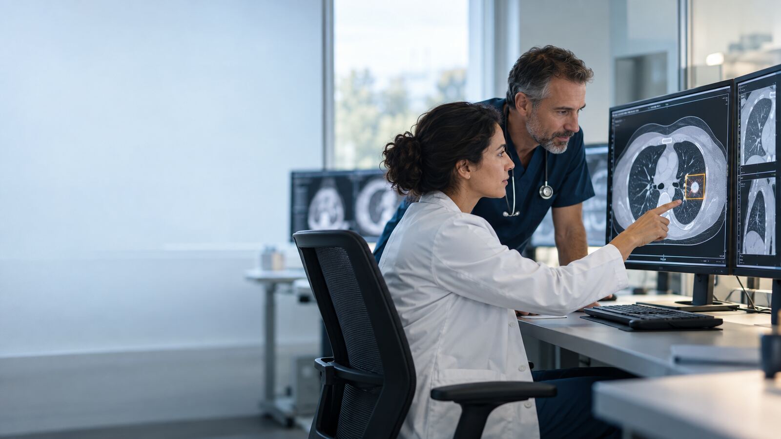
Modern medicine produces more images than any clinician can hold in view at once. X-rays, CT scans, MRIs and ultrasounds move through specialized worklists, reports and electronic records. Radiologists interpret them, while emergency physicians, family doctors, surgeons and other specialists must connect those findings to the patient’s broader story. At every handoff, something important can become less visible.
This is the radiology blind spot: not one single error, but a collection of places where a finding may be missed, delayed, misunderstood or never followed. A subtle abnormality can escape attention. An urgent study can wait behind routine cases. An incidental nodule can be documented but lose its follow-up. A referring clinician can read the report without seeing the image or understanding the radiologist’s level of concern.
The video below introduces how clinical AI can help close these gaps. Properly designed systems can act as a second set of computational eyes, prioritize suspicious studies, structure measurements and track recommendations. But AI does not eliminate the blind spot on its own. The safest model is radiologist plus clinical AI plus a reliable communication and follow-up workflow.
What creates a radiology blind spot?
Diagnostic imaging is information-dense. A single scan may contain hundreds or thousands of images, and the radiologist must search systematically while integrating the clinical question, prior studies and technical quality. Fatigue, interruptions, unusual anatomy, low disease prevalence and competing abnormalities can all affect perception.
Yet the blind spot extends beyond visual interpretation. The examination may arrive without sufficient clinical history. Prior imaging may be stored elsewhere. The report may use cautious language that the treating clinician interprets differently. A recommendation may require action months later, when the original encounter has ended and responsibility is unclear.
Clinical AI can address several parts of this chain because it can analyze images and workflow data consistently at scale. The important word is address, not eliminate. Models have their own blind spots, including false negatives, false positives, dataset bias and sensitivity to changes in scanners or protocols.
| Blind spot | How clinical AI may help | Why human oversight remains necessary |
|---|---|---|
| Urgent study buried in a worklist | Flag a suspected time-sensitive finding for earlier review | Priority flags can be wrong and cannot replace clinical triage |
| Subtle or unexpected abnormality | Mark a region for a second look or provide a measurement | The radiologist must interpret the whole examination and context |
| Incidental finding loses follow-up | Extract the recommendation and track whether action occurred | A clinician must confirm relevance and agree on the plan |
| Report meaning is unclear | Structure key findings and route critical communication | Nuance, uncertainty and patient-specific implications require discussion |
1. Prioritizing the studies that cannot safely wait
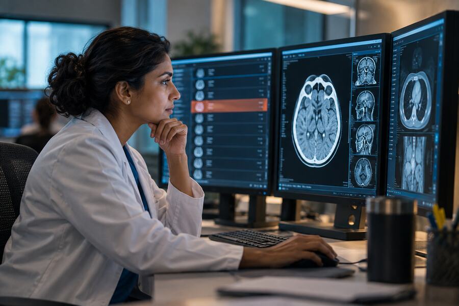
Many radiology departments manage continuous queues from emergency, inpatient and outpatient care. Traditional worklists use factors such as arrival time, location and requested urgency. Those signals are important, but they may not reveal that a newly acquired image contains a potentially time-sensitive abnormality.
A validated clinical AI system can screen incoming studies for a defined finding and move a suspicious case higher in the queue. The system is not issuing the final diagnosis; it is helping direct attention. Earlier review can support faster communication when every minute matters.
This benefit depends on thoughtful integration. If too many routine cases are flagged, radiologists experience alert fatigue and the priority list loses meaning. If the system misses a case, staff must not assume an unflagged examination is normal. The original reading process must remain intact.
Priority algorithms should also account for existing clinical urgency. A scan requested for a deteriorating patient should not be displaced merely because another image triggered a model. The safest design combines clinical triage, acquisition time and AI output in a transparent way.
Performance should be measured using operational outcomes, not only model accuracy. Does the tool reduce time to review for true urgent cases? Does it delay unflagged patients? How often does it generate false alarms? Does the effect remain consistent across shifts, sites and scanners?
Worklist prioritization illustrates the strength of narrow clinical AI. It solves a specific coordination problem and leaves interpretation with the radiologist. Its value comes from the complete workflow—not from an isolated prediction.
2. Providing a second look without creating false certainty
Image-analysis systems can highlight a suspected lesion, identify an asymmetry, estimate a measurement or compare a region across time. Used appropriately, these functions can prompt a radiologist to reconsider an area that might otherwise receive less attention.
The second look is useful because human and model errors are not always identical. A system may detect a pattern that is visually subtle, while the radiologist may recognize contextual clues, artifacts or alternative explanations that the model cannot evaluate. Combining complementary strengths can improve performance when the interface supports independent judgment.
Timing matters. Showing the AI result before the radiologist forms an opinion may increase anchoring: the reader’s attention is directed toward the highlighted region and away from other findings. Showing it after an initial review may preserve independence but add time. The correct design depends on the task and should be tested with intended users.
A bounding box is not an explanation. It may indicate where the system responded without explaining whether the signal reflects pathology, normal anatomy, technical artifact or a feature correlated with the training data. The radiologist must remain responsible for interpreting the whole examination.
| AI assistance | Potential benefit | Possible failure | Required safeguard |
|---|---|---|---|
| Region highlight | Directs attention to a subtle pattern | Anchors the reader or marks an artifact | Independent full-image review |
| Automated measurement | Improves consistency and saves time | Incorrect boundary or wrong structure | Visible contour and editable result |
| Prior comparison | Surfaces interval change | Mismatched studies or protocol differences | Radiologist verifies comparability |
| Drafted impression | Supports completeness and structure | Fabrication, omission or overstatement | Radiologist authors and approves the final report |
3. Closing the incidental-finding follow-up gap
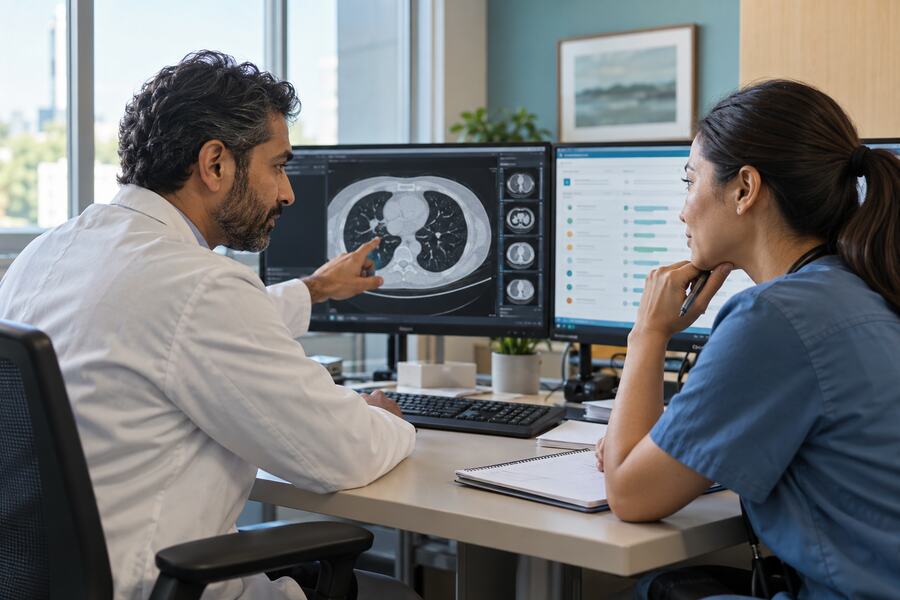
Imaging often reveals an abnormality unrelated to the original reason for the examination. The radiologist may recommend additional imaging, clinical correlation or follow-up after a defined interval. The recommendation enters the report, but the patient’s journey continues across departments and sometimes across organizations.
This creates a coordination blind spot. The ordering clinician may assume another specialist will act. The recommendation may not produce a trackable order. The patient may change providers, miss an appointment or never understand why follow-up matters. A correct interpretation can therefore fail to produce care.
Clinical AI and natural-language processing can help identify recommendation language, convert it into a structured item and connect it to a registry or task queue. The system can check whether the expected action occurred and remind the responsible team when it remains unresolved.
Automation must distinguish a genuine recommendation from general educational wording. It should preserve the radiologist’s level of concern, suggested timing and any uncertainty. A generic reminder without clinical context can create unnecessary testing or fail to communicate urgency.
Responsibility must be explicit. AI can find and route the signal, but an accountable clinician or follow-up team needs to review it, communicate with the patient and determine the appropriate plan. Closing the loop is a clinical and organizational process, not simply a software notification.
Tracking also creates useful quality information. Organizations can measure how often recommendations are acknowledged, scheduled, completed or lost. These data can reveal barriers affecting particular sites or patient groups and support targeted improvement.
4. Translating the report into coordinated care
Radiology reports are written for clinical professionals, but referring doctors have different levels of imaging expertise. Descriptive language, probability terms and recommendation phrases may be interpreted inconsistently. A finding can be technically communicated yet still fail to create a shared understanding.
Clinical AI may help structure key findings, link them to the relevant image, compare the impression with the clinical question and route urgent results through established communication channels. It can also identify when documentation lacks a required acknowledgment.
However, an automated summary must not flatten nuance. “Cannot exclude,” “suspicious for” and “consistent with” carry different meanings. The model should not turn uncertainty into certainty or generate a treatment recommendation beyond the report. Direct communication between clinicians remains essential for unexpected or critical findings.
New blind spots created by clinical AI
Every safety tool can introduce new failure modes. A model may perform differently when a hospital changes scanners, reconstruction software, patient mix or imaging protocols. It may produce uneven results across demographic groups or uncommon conditions. A subtle integration failure can prevent the system from receiving an image without making the absence obvious.
Automation bias may cause users to overvalue a positive result or feel reassured by the absence of a flag. Alert fatigue can make important warnings easier to ignore. If an AI overlay is visually dominant, it may narrow the radiologist’s search rather than extend it.
These risks make local validation and ongoing monitoring essential. Authorization or success at another institution does not prove that performance will remain the same in every environment. The organization must understand intended use, limitations and the conditions under which the model was evaluated.
| Risk signal | What it may indicate | Operational response |
|---|---|---|
| Sudden change in alert frequency | Input, protocol, integration or population shift | Investigate by site, scanner and software version |
| High false-positive burden | Threshold mismatch or poor local fit | Assess alert impact and validated configuration options |
| Low radiologist disagreement | Good performance—or uncritical acceptance | Audit representative cases and review user behaviour |
| Unequal results across subgroups | Data imbalance or hidden bias | Investigate harm, restrict use and require corrective evidence |
| Missed or delayed follow-up tasks | Ownership or workflow failure | Assign responsibility and redesign escalation |
Build a monitored radiologist–AI partnership
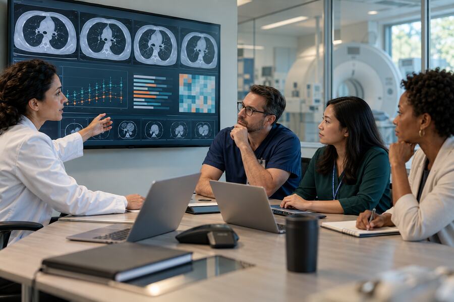
Safe deployment begins with a precise problem. Organizations should identify which blind spot they are addressing, which users and patients are affected, what action the AI may influence and what happens when its output is wrong or unavailable.
Evaluation should use representative local cases and clinically meaningful endpoints. Sensitivity and specificity matter, but so do reading time, turnaround, alert burden, follow-up completion and effects on unflagged studies. The combined radiologist–AI team should be tested rather than the model alone.
Training must include failure cases. Radiologists and referring clinicians should understand the model’s purpose, excluded populations, common false positives, possible false negatives and correct escalation route. Users must know that no alert is not the same as no disease.
The system should preserve independent review and make disagreement easy. Users need to correct measurements, dismiss misleading markers and report suspected failures without interrupting care. Those corrections should support quality review rather than disappear into an unused log.
Monitoring should continue throughout the lifecycle. Performance must be reviewed by scanner, site and relevant subgroup where feasible. Updates to the model, interface, thresholds or integrations should be assessed before release, with rollback procedures available.
Governance should include radiology, referring clinicians, imaging technologists, medical physics, information technology, privacy, quality and patient-safety leaders. Clinical AI affects the entire imaging pathway, so responsibility cannot rest with the algorithm vendor or one enthusiastic department.
A practical readiness checklist
| Area | Question to ask before deployment |
|---|---|
| Purpose | Which exact radiology blind spot does the system address? |
| Evidence | Was performance evaluated for the intended finding, population, modality and setting? |
| Workflow | How does the output change attention, priority, reporting or follow-up? |
| Human control | Can users inspect, disagree, correct and safely continue without the AI? |
| Equity | Are performance differences across relevant groups understood and monitored? |
| Monitoring | Who reviews errors, drift, complaints and operational impact? |
| Accountability | Who owns follow-up when the system identifies a concern? |
Clinical AI should make important findings harder to lose
The greatest promise of clinical AI in radiology is not a machine replacing the specialist. It is a safer information pathway: urgent images rise sooner, subtle regions receive another look, recommendations become trackable and critical findings reach the right person.
That promise is achieved only when technology is connected to responsibility. A model can flag an image, but a trained professional must interpret it. Software can create a follow-up task, but a care team must own it. A dashboard can reveal drift, but leaders must act when performance becomes unacceptable.
Clinical AI does not make radiology infallible. It can make defined blind spots more visible and measurable. Used with validated evidence, thoughtful integration and continuous monitoring, it can help doctors spend attention where it matters while preserving the judgment and communication that safe care requires.
Important: This article provides general educational information and is not medical, legal or regulatory advice. AI-supported imaging tools must be used only within their intended purpose and applicable professional, organizational and regulatory requirements. AI output does not replace interpretation by qualified healthcare professionals or emergency care.
Further reading
| Organization | Resource | Focus |
|---|---|---|
| World Health Organization | Ethics and governance of artificial intelligence for health | Human autonomy, safety, transparency, accountability and equity |
| World Health Organization | Ethics and medical radiological imaging | Ethical issues surrounding AI and radiological imaging |
| Health Canada, FDA and MHRA | Good machine learning practice for medical device development | Representative data, human–AI performance and lifecycle management |